Catalan researchers create technique to remove brain tumors without affecting language processing
Study applies magnetic resonance in the inferior fronto-occipital fascicle of cerebrum
Research led by neuroradiologists, who specialize in the nervous system, from the Catalan Bellvitge hospital has removed brain tumors without affecting the inferior fronto-occipital fasciculus (IFOF) of the cerebrum, which is associated with semantic language processing and goal-oriented behavior.
The study has been conducted in association with the Institute of Diagnostic Imaging (IDI), and the Biomedical Research Institute of Bellvitge (IDIBELL).
Preserving the IFOF area of the brain during surgery is an "essential" factor to avoid permanent neurological damage and has a huge impact on the patients’ quality of life.
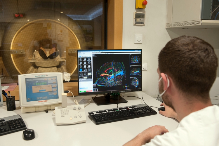
Researchers have undertaken the study based on an advanced magnetic resonance techniques program that focuses on the inferior fronto-occipital fasciculus.
Using the diffusion tensor method, professionals can precisely locate and determine the size of a tumor without affecting the IFOF. If the area is in fact injured, the patient can even deform and replace words while speaking.
"Surgeons will be able to systematically identify with a non-invasive operation the fasciculus, helping to precisely defy the limits to remove a brain tumor," doctor Àngels Camins, leader of the project, explained.
The Bellvitge hospital started its studies on language with magnetic resonance around 12 years ago. Its goal was to bring to light the best pre-surgery plan possible for patients with tumors which have a high risk of suffering post-operation deficiencies.